Home » Without Label » Anatomy Diagram Rib Area - Human Rib Cage Photos And Premium High Res Pictures Getty Images : The top thoracic vertebra, t1, connects with c7 in the cervical spine above while the bottom thoracic vertebra, t12, connects with l1 in the lumbar spine below.
Anatomy Diagram Rib Area - Human Rib Cage Photos And Premium High Res Pictures Getty Images : The top thoracic vertebra, t1, connects with c7 in the cervical spine above while the bottom thoracic vertebra, t12, connects with l1 in the lumbar spine below.
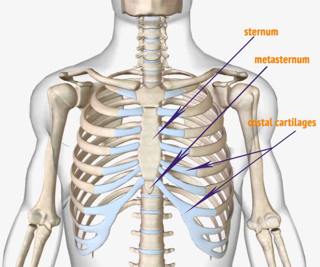
Anatomy Diagram Rib Area - Human Rib Cage Photos And Premium High Res Pictures Getty Images : The top thoracic vertebra, t1, connects with c7 in the cervical spine above while the bottom thoracic vertebra, t12, connects with l1 in the lumbar spine below.. The sternum is located in the center of the anterior thoracic wall and is also known as the breastbone. The sternum is a flat bone that is made up of three parts, the (1) manubrium, (2) body, and the (3) xiphoid process. It is a large depressed area that lies near the centre of the medial surface. This anatomical module is about the anatomy of the mediastinum and the sectional anatomy of the thorax. Count the ribs and intercostal spaces.
The muscles of the abdomen protect vital organs underneath and provide structure for the spine. Gross anatomy of lungs 2. Anterior end of 1st rib.rib. Hilum and root of lungs 4. It has a roughened area on its upper surface, from which the serratus anterior muscle originates.
Anatomy Of The Human Ribs With Full Gallery Pictures Dislocated Rib from dislocatedrib.org Its functions are to protect the thoracic organs from trauma and also form the bony attachment for various muscles. The typical rib consists of a head neck and body. The anatomy of the human ribs (costae) are one of the integral parts of the chest wall; The rib cage is the arrangement of ribs attached to the vertebral column and sternum in the thorax of most vertebrates that encloses and protects the vital organs such as the heart, lungs and great vessels. Ribs 11 and 12 do not have necks or tubercles and the anterior tips of. Rib cage, in vertebrate anatomy, basketlike skeletal structure that forms the chest, or thorax, and is made up of the ribs and their corresponding attachments to the sternum (breastbone) and the vertebral column.the rib cage surrounds the lungs and the heart, serving as an important means of bony protection for these vital organs.in total, the rib cage consists of the 12 thoracic vertebrae and. Our latest youtube film is ready to run. The rib cage is an important part of the human anatomy.
The major muscles of the abdomen include the rectus.
Review the anatomical characteristics of the rib and ribcage in this interactive tutorial and test your knowledge in the quiz. These pains may be stemming from the right or. Sternum and ribs diagram quizlet. The typical ribs have a generalised structure while the atypical ribs have variations on this structure. The heads of ribs 1, 10, 11, and 12 have a single facet for articulation with the bodies of the thoracic vertebrae. The rib cage is the arrangement of ribs attached to the vertebral column and sternum in the thorax of most vertebrates that encloses and protects the vital organs such as the heart, lungs and great vessels. The major muscles of the abdomen include the rectus. The thoracic spine is comprised of 12 vertebrae labeled t1 through t12. Our latest youtube film is ready to run. Several muscles that move the arms, head, and neck have their origins on the sternum. This anatomical module is about the anatomy of the mediastinum and the sectional anatomy of the thorax. Surfaces and borders of lungs 3. The rib cage is an important part of the human anatomy.
Genderless torso with sternum and ribs 16 part. The thoracic spine is comprised of 12 vertebrae labeled t1 through t12. Human rib cage anatomy diagram. Several muscles that move the arms, head, and neck have their origins on the sternum. Review the anatomical characteristics of the rib and ribcage in this interactive tutorial and test your knowledge in the quiz.
Rib Cage Organ Thoracic Cavity Internal Thoracic Artery Organs Heart Lung Anatomy Png Pngwing from w7.pngwing.com We are pleased to provide you with the picture named heart, lung, diaphragm and ribs location.we hope this picture heart, lung, diaphragm and ribs location can help you study and research. Rib 2 is thinner and longer than rib 1, and has two articular facets on the head as normal. In addition to being connected to adjacent vertebrae, the thoracic vertebrae. 11 best photos of posterior ribs diagram displaced 122 123a rib from the middle of the series should be taken in order to study the common characteristics of these bones. This anatomical module is about the anatomy of the mediastinum and the sectional anatomy of the thorax. Each pair is numbered based on their attachment to the sternum, a bony process at the front of the rib cage which serves as an anchor point. Rib 1 is also flattened horizontally. Several muscles that move the arms, head, and neck have their origins on the sternum.
They make up the lateral part of our body, its anterior and posterior wall and they entirely build the lateral parts of the chest wall.
Count the ribs and intercostal spaces. The typical ribs have a generalised structure while the atypical ribs have variations on this structure. Female anatomy includes the external genitals, or the vulva, and the internal reproductive organs. Vital organs such as heart and lungs are protected by the rib cage. These pains may be stemming from the right or. Our latest youtube film is ready to run. Gross anatomy of lungs 2. The muscles of the abdomen protect vital organs underneath and provide structure for the spine. They make up the lateral part of our body, its anterior and posterior wall and they entirely build the lateral parts of the chest wall. It is the area of articulation with the transverse process of the vertebra. Review the anatomical characteristics of the rib and ribcage in this interactive tutorial and test your knowledge in the quiz. The bones of the rib cage are the sternum, the 12 thoracic vertebrae and the 12 pairs of ribs. The top thoracic vertebra, t1, connects with c7 in the cervical spine above while the bottom thoracic vertebra, t12, connects with l1 in the lumbar spine below.
The part of the muscle is thought. True ribs, false and floating. Female anatomy includes the external genitals, or the vulva, and the internal reproductive organs. The major muscles of the abdomen include the rectus. Anterior end of 1st rib.rib.
Anatomy Color Coded Lungs Inside Rib Cage 3d Illustration Stock Photo Download Image Now Istock from media.istockphoto.com Rib 2 is thinner and longer than rib 1, and has two articular facets on the head as normal. Vital organs such as heart and lungs are protected by the rib cage. The thoracic spine is comprised of 12 vertebrae labeled t1 through t12. In addition to being connected to adjacent vertebrae, the thoracic vertebrae. The sternum, commonly known as the breastbone, is a long, narrow flat bone that serves as the keystone of the rib cage and stabilizes the thoracic skeleton. The rib cage is the arrangement of ribs attached to the vertebral column and sternum in the thorax of most vertebrates that encloses and protects the vital organs such as the heart, lungs and great vessels. Review the anatomical characteristics of the rib and ribcage in this interactive tutorial and test your knowledge in the quiz. Ribs 11 and 12 do not have necks or tubercles and the anterior tips of.
Related posts of rib cage diagram with organs womens body parts stomach.
Gross anatomy of lungs 2. These muscles help the body bend at the waist. Anatomy of the rib cage diagram anatomy of the rib cage diagram in this image, you will find thoracic vertebrum, costochondral joint, costal cartilage, costal margin, costal arch, thoracic vertebrum, xiphoid process, xiphisternal joint, body, manubrial sternal joint, manubrium, the sternal notch in it. Human ribs drawing at getdrawings com free for personal. The rib cage is a bony structure found in the chest (thoracic cavity). Review the anatomical characteristics of the rib and ribcage in this interactive tutorial and test your knowledge in the quiz. Anatomical atlas of the mediastinum and thorax : The thoracic spine is comprised of 12 vertebrae labeled t1 through t12. It is made up of 12 pairs of ribs. The sternum is a flat bone that is made up of three parts, the (1) manubrium, (2) body, and the (3) xiphoid process. Related posts of rib cage diagram with organs womens body parts stomach. True ribs, false and floating. Genderless torso with sternum and ribs 16 part.